In ultrasound imaging, 3D and 4D imaging is a relatively new technique. In 3D ultrasound imaging, several 2D slice captures are made and digitally preserved. These can then result in lifelike images. Simply put, 4D scanning allows for the real-time viewing of these genuine images, allowing for the study of the baby's movements.
No matter how many times we view it, we can never help but feel the same thrill that parents have when they see their unborn child for the first time on the ultrasound screen! It's really exciting to see them when they experience their child's rapid pulse for the first time!
The main reason parents want an ultrasound is to feel the excitement of the procedure, but the true purpose is to ensure the baby is healthy and developing correctly. The number of ultrasounds a pregnant woman has depends on several variables, including the specific OB/GYN's practice, whether or not the pregnancy is uncomplicated and healthy, and so on.
Although ultrasounds are safe, more than what is medically necessary should not be done. Having said that, there are numerous reasons why having an ultrasound performed is a crucial component of your prenatal care.
Benefits of 3d and 4d Ultrasound
It’s good for the incredible progress in both technology and medicine, 3d-4d imaging ultrasound in Sacramento can now provide unprecedented levels of information! This has made it much easier for us to evaluate the baby's well-being.
Additionally, it has made it possible for medical professionals to see more minute alterations linked to certain pregnancy-related issues. The following are some benefits of 3D and 4D ultrasounds and how they can be used to find vital information on the health of your unborn child:
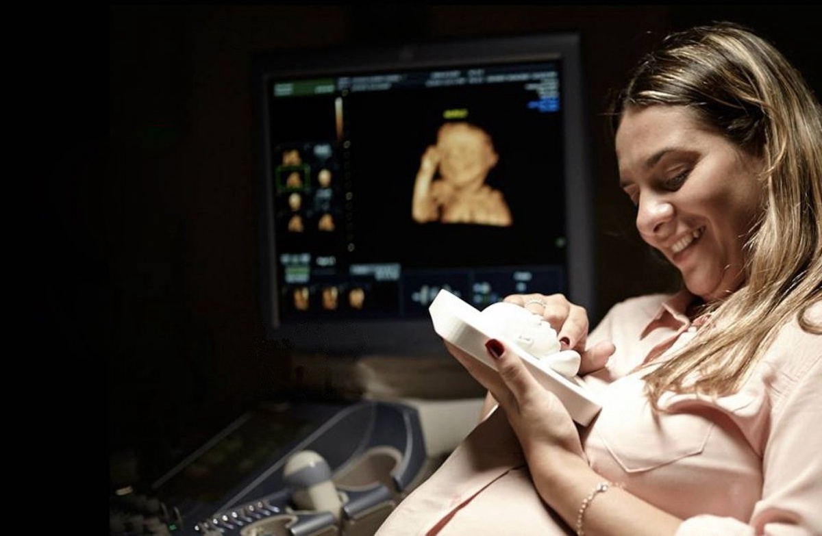
Ultrasound photo creation technology has advanced significantly, and these images are frequently the first you add to your scrapbook. Instead of merely having a small black-and-white picture on the refrigerator as your parents did, you can decorate yours with a highly detailed portrait of your unborn child taken during a 4D ultrasound.
Our innovative ultrasound equipment uses contemporary technology to provide 3d-4d imaging ultrasound in Sacramento of the child. Anyone can view the unborn child's entire face, down to the smallest facial detail, as well as all of its internal movements, such as kicking, giggling, and thumb-sucking.
This not only allows you to bond from the start, but it also assures you that your baby appears healthy. If your pregnancy is difficult, a 3D and 4D ultrasound can help to achieve the best possible conclusion.
Functions of ultrasounds
The differences between a 4D and a regular ultrasound are minimal. The ultrasonography machine detects the sound waves that bounce back after we send them through the womb. A 3d-4d imaging ultrasound in Sacramento has the benefit of taking images from various perspectives. These pictures can be merged to make a detailed, moving picture.
The advantages of 3d-4d ultrasonography
The ability to see your unborn child before they are born is undoubtedly the draw of an ultrasound. Any excited parent will find it exciting to see their baby's facial characteristics and see them move in real-time during a 4D ultrasound.
Both you and your child can feel safe and comfortable using ultrasound technology. Sound wave technology is used in both 3D and 4D ultrasounds to produce an image of the target location, which can be breast and uterine tissues or a baby in the womb.
One of the primary advantages of obtaining a 3D or 4D ultrasound is that it can assist our team in identifying potential health problems and birth deformities that might not be evident in other imaging procedures.
Based on the stage of fetal development, you might also be able to obtain a more comprehensive image of your baby's face and characteristics. Still, there are many advantages to having a thorough ultrasound. Because 4D ultrasounds create substantially clearer images, they can be utilized to determine birth abnormalities.
Little deformities called cleft lips and palates are frequently missed by 2D ultrasounds but are visible with 3D/4D imaging. This allows medical professionals to inform parents about the illness and help them prepare for any complications.
Affordable 3d-4d ultrasound in Sacramento can help put individuals in touch with a neonatologist who can help you through the process of creating a treatment plan for your child if they have a birth defect that is visible in 4D. The normal and abnormal fetal anatomy can be shown in controlled planes and rendered images from various perspectives thanks to the availability of many display modes and standardized testing.
This makes it possible to display even minute fetal abnormalities in the perfect sectional plane on a transparent image or accurately rendered surface when seen from the best possible perspective. The produced images can be used to reassure parents that there is no fetal abnormality present or, conversely, to help them grasp the severity of an existent deformity while counseling the parents. This is especially helpful when there is a higher chance of a certain fetal abnormality recurring.
Conclusion
Over the past three decades, there has been a significant advancement in echocardiography, moving from M-mode imaging to 3d-4d imaging and finally to the developing role of 3d and 4d imaging modalities.
The examination of numerous cardiac conditions has greatly benefited from 3d echocardiography, which has also become a crucial component of standard clinical practice and the basis for clinical recommendations for the treatment of heart disease.
Nevertheless, it is well known for having several drawbacks, such as the absence of 4d spatial dimensions, apical foreshortening, geometric assumptions, and poor boundary identification. Consequently, it became clear that 4d visualization and analysis would be a more effective way to represent and comprehend the intricate 4d architecture and physiological processes of the human body.
It is projected that as 3d and 4d technology advances through advances in transducer technology, picture quality, standardization, and software development, 3d and 4d will gradually find its way into clinical guidelines and everyday practice.

No comments yet